본문바로가기

SoniX EchoPilot LVH™

(MFDS Class III Approval)

Differential Diagnosis Support.
Assists clinicians in differentiating
the underlying causes of myocardial hypertrophy,
including hypertrophic cardiomyopathy (HCM),
cardiac amyloidosis, and hypertensive heart disease.

SONIX HEALTH
SCROLL DOWN
The Challenge
Diagnostic Challenges
in Cardiomegaly Etiology

Conventional echocardiography imaging alone
makes it exceptionally difficult to distinguish
the precise underlying causes of myocardial hypertrophy.

For instance, the diagnostic sensitivity
for hypertensive heart disease stands at a mere 33%,
leaving a significant number of patients undiagnosed.

  • Limitations of Visual Differentiation

    Because various underlying causes of myocardial hypertrophy present highly similar echocardiographic features, distinguishing them by visual inspection alone is exceptionally difficult.

  • Low Diagnostic Sensitivity

    For instance, when relying solely on conventional visual assessment, the diagnostic sensitivity for hypertensive heart disease is a mere 33%.

  • Challenges in Differential Diagnosis

    Achieving a definitive diagnosis often requires expensive additional imaging such as CT or MRI, or even invasive biopsies. This dependency can ultimately delay timely and appropriate treatment intervention.
The Process

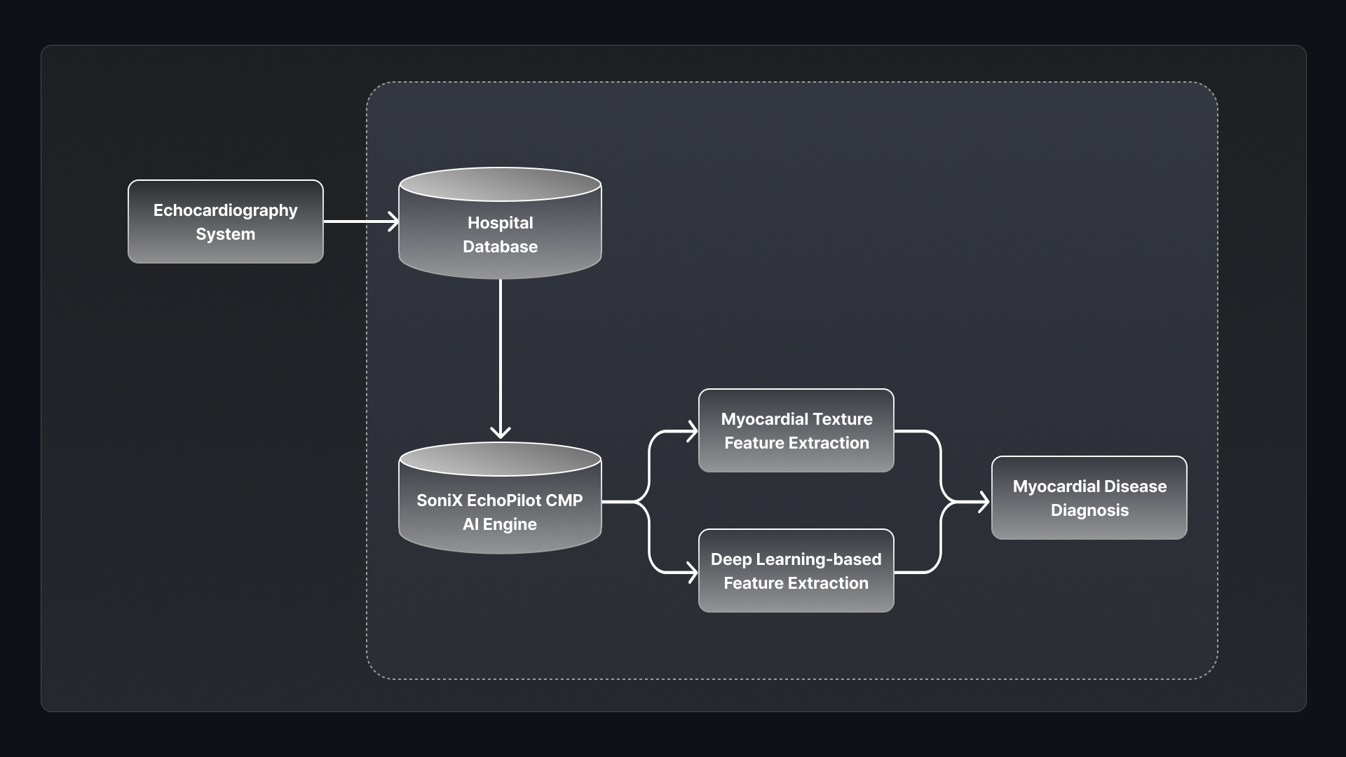
SoniX EchoPilot LVH™ Workflow

Core Features
An AI-driven solution
assisting in the differential diagnosis
of myocardial hypertrophy.
AI Screening Solution
for Myocardial Hypertrophy
Differential Diagnosis.

SoniX EchoPilot LVH™ is a primary AI screening solution
that automatically classifies the underlying causes
of myocardial hypertrophy using echocardiography images alone.

It performs primary screening for the risk of major,
clinically challenging conditions-including
Hypertrophic Cardiomyopathy (HCM), Cardiac Amyloidosis,
and Hypertensive Heart Disease-using echocardiography images alone,
eliminating the need for expensive follow-up tests like CT or MRI.

※ Clinical Evidence: Artificial Intelligence-Enhanced Analysis of
Echocardiography-Based Radiomic Features for Myocardial Hypertrophy
Detection and Etiology Differentiation (2025)

5 Steps
of AI Analysis
  • Step 1: Image Quality Assessment
    During the image input stage, the system automatically evaluates the quality of left ventricular (LV) echocardiography images to determine their suitability for analysis, automatically excluding non-standard views.
  • Step 2: Cardiac Structure Segmentation
    The segmentation AI automatically extracts left ventricular (LV) myocardial borders to secure the entire myocardial area, serving as the essential foundation for highly accurate measurements.
  • Step 3: Myocardial Feature Extraction
    The system extracts key characteristics such as myocardial shape, thickness, and texture. This data serves as critical information for accurate differential diagnosis.
  • Step 4: Pattern Analysis
    The system calculates the probability of each condition by comparing the three extracted features against trained disease patterns. It achieves exceptional accuracy through complex, multi-dimensional pattern recognition.
  • Step 5: Risk Assessment and Reporting
    The system provides risk assessments for major conditions and detailed screening reports to support rapid diagnostic and treatment workflows.
Performance of SoniX EchoPilot LVH™
Differential Diagnosis Performance
for the 3 Major Causes
of Myocardial Hypertrophy
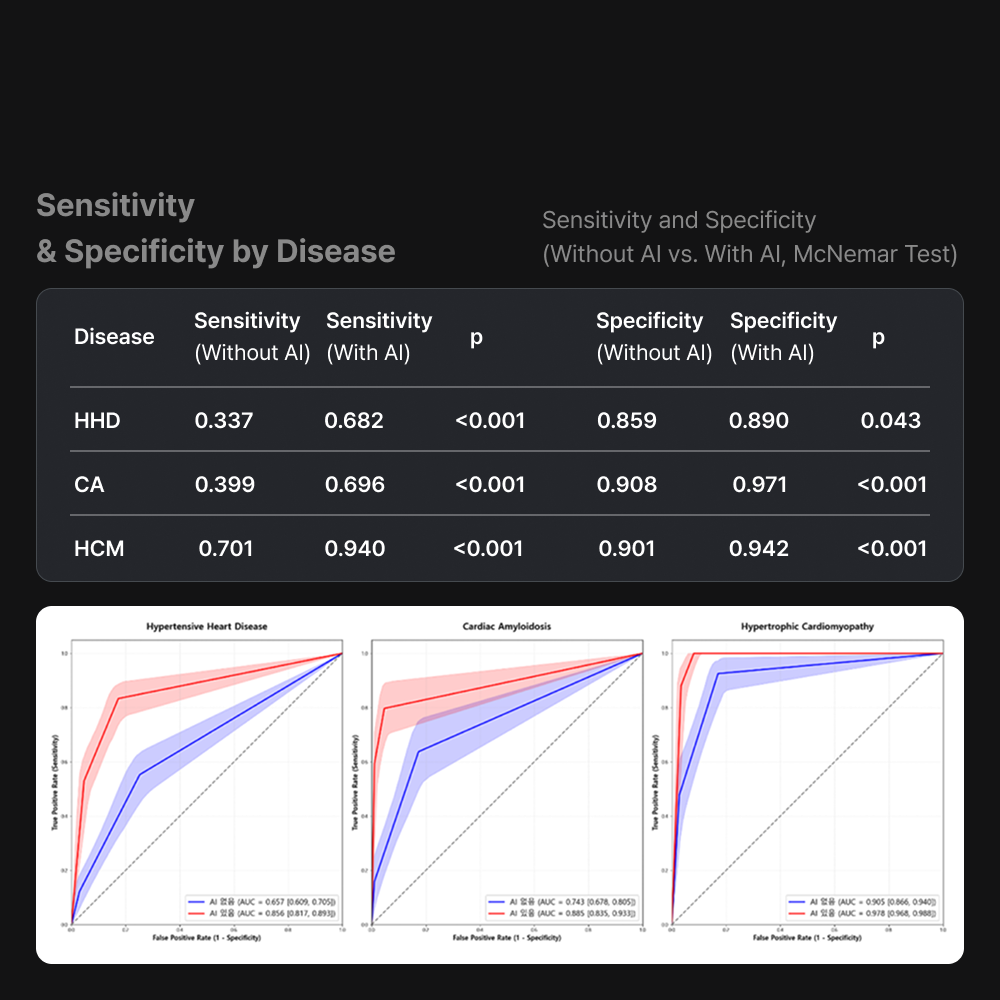
제품 성능 표
Category Hypertrophic Cardiomyopathy (HCM) Cardiac Amyloidosis Hypertensive Heart Disease
Clinical Characteristics Genetic, asymmetric hypertrophy Protein deposition, restrictive cardiomyopathy Chronic pressure overload, symmetric hypertrophy
Diagnostic Difficulty ★★★ High ★★★ High ★★★★ Very High
Human Diagnostic Sensitivity - - 33%
AI Diagnostic Accuracy 96% 89% 86%
Sensitivity Improvement (AI) - - 33% → 75% (↑ 42%p)
Clinical Significance Prevents sudden cardiac death via early detection Improves prognosis through timely treatment Reduces unnecessary rule-out/exclusionary tests
Three Core Values

SoniX EchoPilot LVH™Engine sets a new standard
for echocardiography interpretation through clinical accuracy,
workflow efficiency, and data standardization.

  • Primary Care Integration

    It integrates seamlessly into existing echocardiography workflows without the need for expensive CT or MRI scans. Its vendor-neutral design significantly lowers the barrier to entry, allowing for immediate adoption at the clinical site.

  • Accelerating Time-to-Treatment

    By enabling early screening for critical conditions that demand urgent intervention—such as hypertrophic cardiomyopathy (HCM) and cardiac amyloidosis—the solution significantly shortens the time to specialist confirmation and treatment initiation.

  • Automated Report Generation

    The system automatically generates a diagnostic report including condition-specific risk levels and clinical recommendations based on the screening results, providing structured support for clinical decision-making.

※ Regulatory Notice: MFDS Class III Approved (License No. D 제허 25-15호) | Undergoing Integrated Review for Innovative Medical Devices
HCM: Hypertrophic Cardiomyopathy | LVH: Left Ventricular Hypertrophy | HD: Heart Disease

This product is a medical device. Please carefully read
and thoroughly understand the 'Precautions
for Use' and 'Instructions for Use' before operation.

Other products
SoniΦ Assist™
SoniΦ Assist™

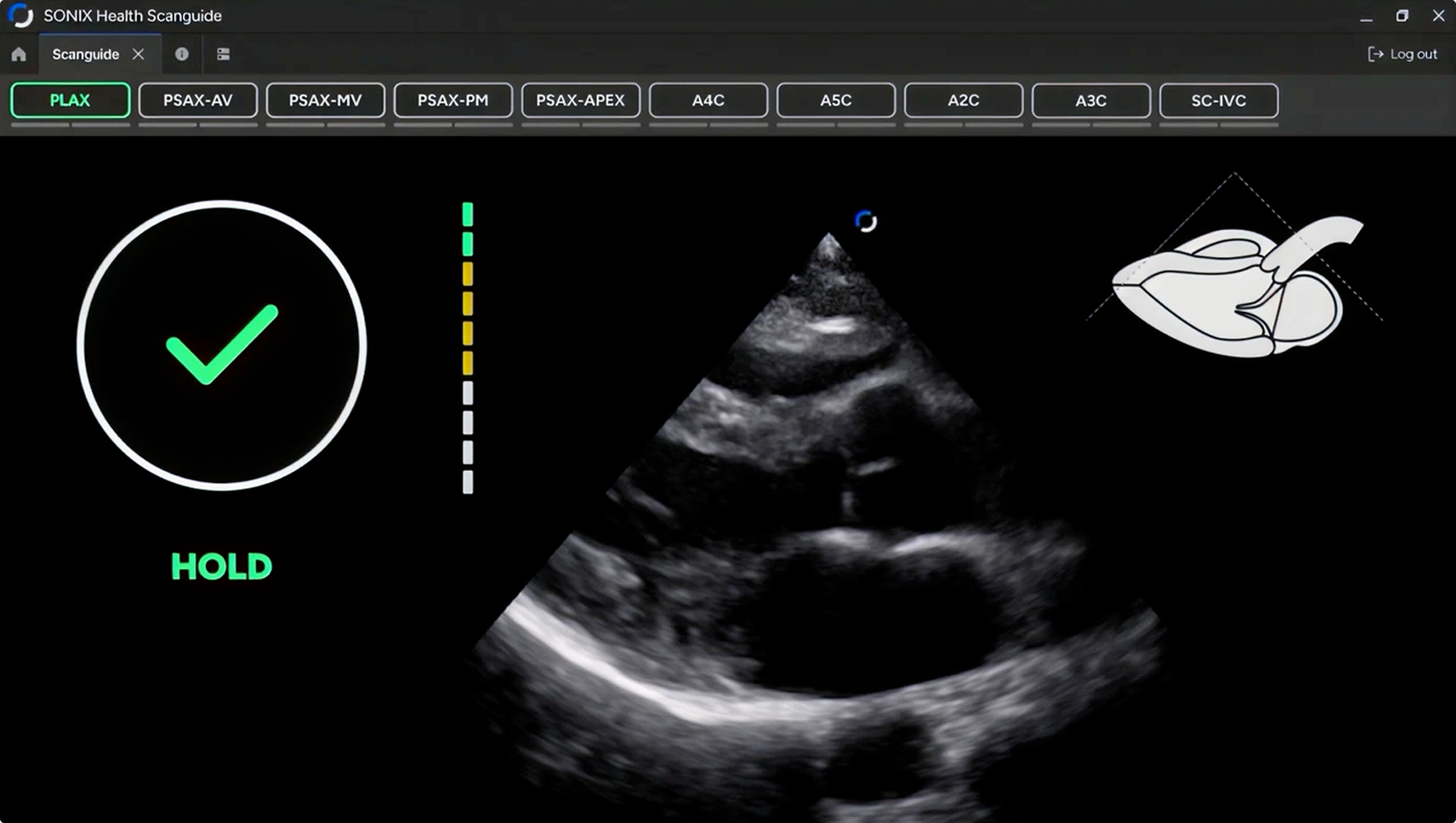
AI Scan Guide.
Provides real-time guidance
to ensure standardized
and high-quality image acquisition.

More View
SoniX EchoQuant™
SoniX EchoQuant™

Automatically measures 81 indicators
across multiple cardiac cycles,
reducing measurement variability
and shortening exam time.

More View

SITEMAP

DRAG